Cytogenetcs
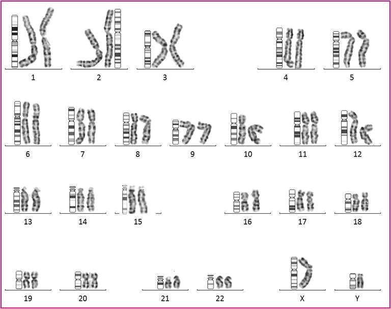
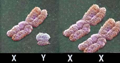
KARYOTYPE Human genome consists of 23 chromosomal pairs: 22 autosomes and one pair of sex chromosomes. It is estimated that the haploid of human genome consist of three billion nucletiotides organized into 23,000 protein-coding genes. About 1.5% of the total human DNA represents coding sequences of the structural and regulatory genes. The rest are non-coding RNA genes, regulatory sequences and evolutionary trash of the DNA. Human karyotype comprises 46 chromosomes (23 chromosomal pairs), 22 autosomes and one pair of sex chromosomes (XX in woman, XY in man).
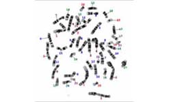
Commonly used methods for karyotyping include the classic cytogenetic technique in combination with GTG (Giemsa-Trypsin-Giemsa) or FISH (fluorescence in situ hybridization). In adults, the most frequent is karyotyping from a blood sample, rarely from skin biopsy, whereas prenatal analyses are performed from chorionic villus, amniotic fluid or cord blood. Karyotyping provides information on the chromosomal structure that consists of several million of nucleotide base-pairs.
Karyotype - prenatal a) Karyotype from amniotic fluid Amniocentesis is a procedure n which amniotic fluid is sampled from the uterus for testing or treatment. Amniocentesis is performed between the 15th-20th weeks of pregnancy. Amniotic fluid is the fluid that surrounds and protects a baby during pregnancy. This fluid contains fetal cells and various chemicals produced by the baby. The procedure is applied in pregnancies where high risk of chromosomopaties is estimated using screening tests such as the first trimester screen and in pregnancies with positive previous history for chromosomopaties as Down syndrome or neural tube defect. The test is also applied if the mother is older than 35, because babies born to women aged 35 and older have a higher risk of chromosomal abnormalities, such as Down syndrome. Amniocentesis is also performed if a positive family history of a specific genetic disorder exists, such as spinal muscle atrophy or cystic fibrosis. The sampling is carried out in gynaecological ordinations or clinics by specialist gynaecologists using ultrasound. Usually 15 ml of amniotic fluid is taken for analysis. Harvesting and analysis are carried out in laboratory: foetal cells are collected and set up into the cell cultures. When the appropriate number of cells in division (which usually takes 2 weeks) is reached, the sample is analyzed under the microscope. By karyotyping we analyze the number and the structure of chromosomes, as well as baby’s gender. In prenatal diagnostics it is sometimes necessary to combine cytogenetic and molecular methods, i.e. the analysis of particular genes or markers. We most commonly combine the screening of STR markers located in chromosomes that are most frequently found in aneuploidies (chromosomes 21, 13, 18 and sex chromosomes) and the karyotype analysis. This approach provides screening results within 24hours, whereas the karyotype analysis is completed within 2 weeks. b) Karyotype from Chorionic villus Chorionic villus sampling (CVS) is a prenatal test in which a sample of chorionic villi is removed from the placenta for testing. Chorionic villus sampling can reveal a kryotype, i.e. whether a baby has a chromosomal abnormality, such as Down syndrome. Chorionic villus sampling can also be used to test for other genetic disorders. For karyotyping a short term cultivation of villus, direct karyotyping, as well as a combination of karyotyping with other techniques for prenatal diagnostics are performed. Chorionic villus sampling is usually done between the 10th and 12th weeks of pregnancy - earlier than other prenatal diagnostic tests, such as amniocentesis. c) Cordocentesis Cordocentesis is a prenatal test in which a foetal blood sample from the umbilical cord is used for genetic testing. Cordocentesis can be performed after 20 weeks of pregnancy. Testing is performed because of genetic problems or infections. Results are usually available within 72 hours. a) Karyotyping in adults is usually performed to find out whether there are extra, missing, or abnormal positions of chromosome pieces that could cause problems with a person's growth, development, and body functions. By karyotyping we determine whether the chromosomes of an adult have an abnormality that can be passed on to a child, whether chromosome rearrangements could be the cause of a miscarriage, or to identify the sex of a person. In particular cases, karyotype can help determine the appropriate treatment for some types of cancer. Results of karyotyping are obtained within 3-5 days. For the analysis a blood sample taken on heparin is used. b) In particular cases, karyotype may be analyzed from skin biopsies. For that purposes, the skin sample 1x1 mm is taken under aseptic conditions, and should be immediately put into sterile physiological saline. Karyotyping from skin takes 10-14 days.
|
|